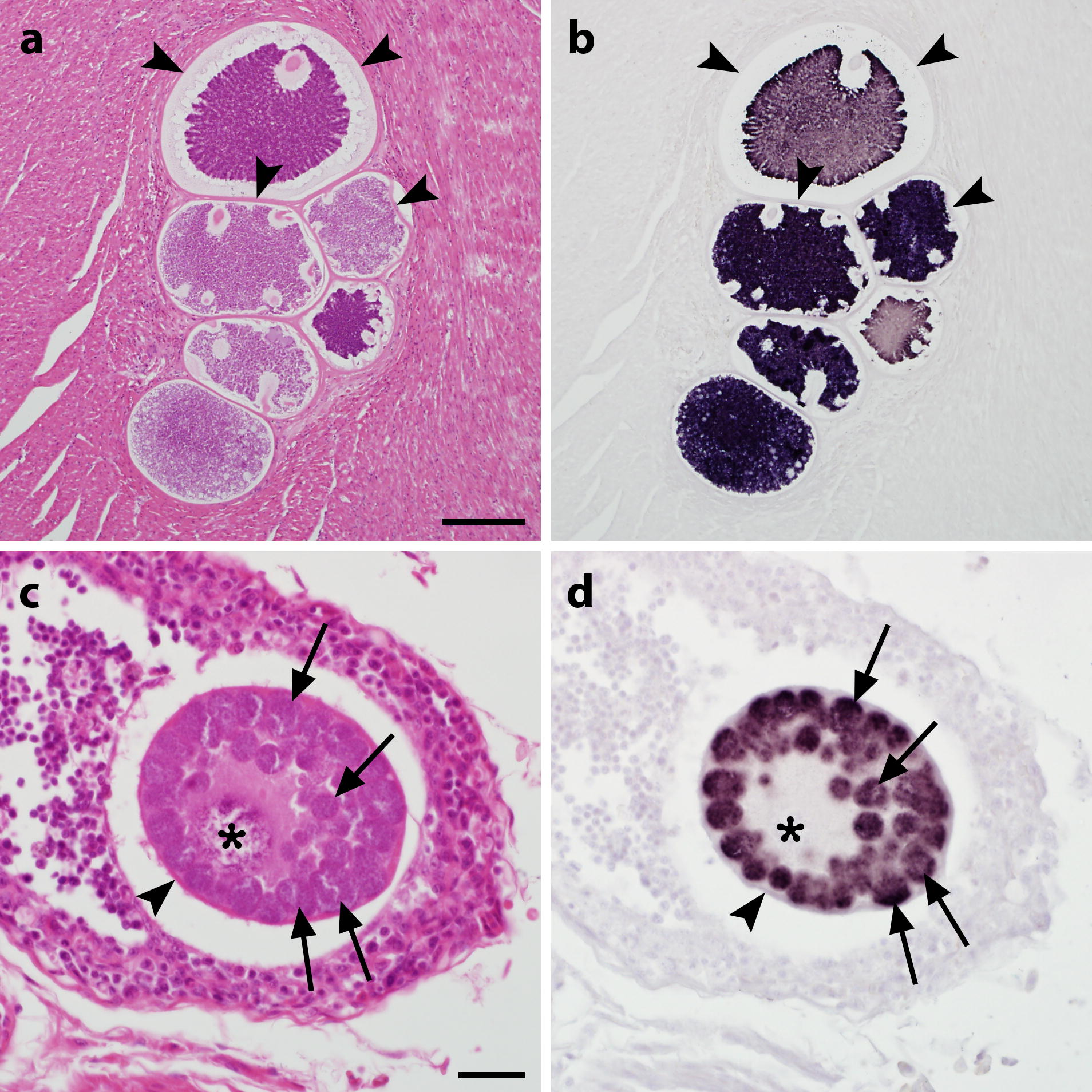
Fig. 2.

Megalomeronts of Haemoproteus minutus (a, b) and Leucocytocoon sp. (c, d) in histological sections from infected parakeets stained by haematoxylin and eosin (HE; a, c) and chromogenic in situ hybridization (CISH; b, d). Multiple megalomeronts of H. minutus, lineage TUPHI01, and Leucocytozoon cf. californicus, lineage CIAE02, were observed in HE-stained sections of the cardiac muscle in a red-crowned parakeet (Cyanoramphus novaezelandiae; a) and in the bursa of Fabricius in a blue-winged parakeet (Brotogeris cyanoptera; c), respectively. Application of subgenus-specific probes for Haemoproteus (Parahaemoproteus) spp. (b) and Leucocytozoon (Leucocytozoon) spp. (d) with CISH unequivocally indicated generic identity of parasites. Signals were confined to cytomeres (arrows) and merozoites of the parasites whereas structures not attributed to the parasite such as host cell nucleus (asterisk) and prominent capsule-like walls (arrowheads) around megalomeronts remained negative. Note numerous roundish cytomeres (arrows) and a readily visible host cell nucleus (or ‘central body’) present in the megalomeront of Leucocytozoon sp., but absent in megalomeronts of Haemoproteus parasites. Scale-bars: a, 100 µm; c, 20 µm
