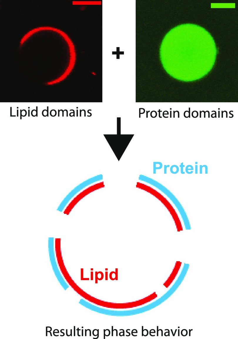
Figure 1.
Reconstitution of interaction between lipid and protein domains. The upper left panel shows a typical confocal fluorescence image of a phase-separated ternary mixture GUV. The signal was from TR-DHPE lipid. The upper right panel shows a typical confocal fluorescence image of a solution phase-separated protein droplet domain from a two-component system (SH3 × 4 and PRM × 4). The signal was from SH3 × 4-Atto488. Scale bars are 5 μm. The lower schematic is a possible outcome of coexistence of both on the lipid bilayer.

