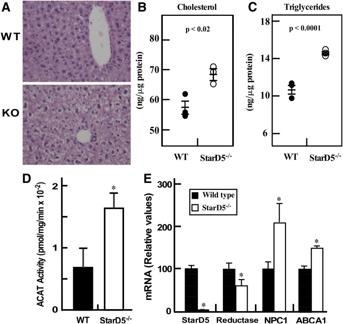
Fig. 3.
StarD5 deletion increases liver lipid content. A: Formalin-fixed sections from WT and StarD5−/− mouse livers were stained with H&E and visualized with a 40× objective using a Nikon Ti-U inverted microscope. Representative images from three mice are shown. (B and C) Liver total cholesterol (B) and triglycerides (C) were quantified as described in the Materials and Methods (n = 3). (D) Microsomes were prepared from WT and StarD5−/− livers and ACAT activity was quantified as described in the Materials and Methods (n = 3, P < 0.05). E: Total RNA was extracted from WT and StarD5−/− mouse livers and used in qRT-PCR as described in the Materials and Methods section, n = 3, *P < 0.05, WT compared with StarD5−/−.

