Fig. 3.
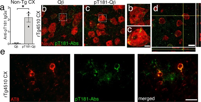
pT181-Qß generated antibodies enter the brain and closely-associate with neurons, positive for pathological tau. Confocal image showing anti-pT181 IgGs were significantly elevated in the cortical lysate of pT181-Qβ vaccinated Non-Tg a. The anti-pT181 IgGs, readily detectable in the pT181-Qβ, but not Qβ control, vaccinated rTg4510 brains (b, c, magnified images of b and c; Streptavidin-conjugated Alexa Fluro 488 detects pT181 antibodies, in green; NeuN, neurons, in red). Orthogonal images, obtained from the confocal microscope, from immunized mice revealed that pT181 antibodies did not colocalize with NeuN, but were likely in the neuronal cytoplasm, as well as in the brain parenchyma d. Epifluorescence images showing anti pT181 IgGs also co-localized to neurons containing pathological tau (AT8) e; Streptavidin-conjugated Alexa Fluro 488 detects pT181 antibodies, in green; AT8, red). Graph displays geometric mean ± 95% Confidence Interval, significance value was determined with a student’s t-test, (p ≤ 0.5 *). Scale bars in b–d are 10 μm; magnified images of b, c are 2 μm, scale bar in e is 20 μm

