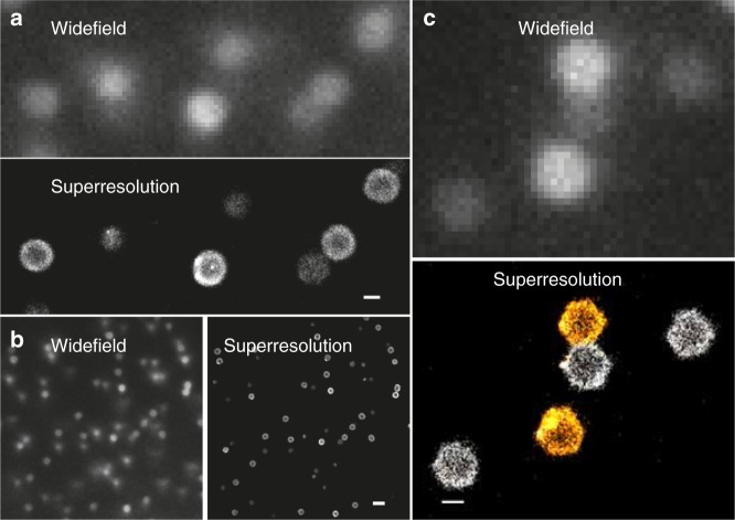
Fig. 1.
Comparison of conventional widefield and dSTORM superresolution images of densely packed microgels. a, b show images of Alexa Fluor®647 dye-labeled microgels seeded in a dense suspension (ζ = 0.86, c = 10.8 wt%). Images are taken in the bulk and thus some of the particles are out of focus and located deeper inside the sample, appearing less bright. This also affects the superresolution imaging leading to a reduced number of localized points that can be used for the reconstruction. c Widefield and two-color dSTORM images of microgels at the glass-sample interface including a pair in contact (ζ = 1.89, c = 23.6 wt%). The widefield image was taken with a low pass filter for Alexa Fluor®647 and thus the microgel labeled with second dye CF680R is barely visible. The two-color image was reconstructed using the spectral demixing technique37, 63. Scale bar 500 nm in panel a and c and 2 μm in panel b

