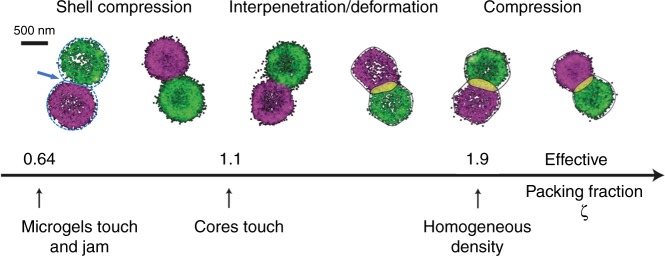
Fig. 2.
Two-color dSTORM superresolution microscopy of microgel pairs. Examples for dye-labeled tracer particles seeded in densely packed microgel suspensions are shown (T = 22 °C)37. Particles are labeled with the fluorophores Alexa Fluor®647 or CF680R. The effective packing fraction ζ increases from left to right (ζ = 0.86, 1.01, 1.26, 1.50, 1.89, 2.13). Left: The dashed circles with radius Rtot = 470 nm visualize the total microgel size including the barely visible low-density corona. The arrow points to the contact area where the brush-like corona is partially compressed. The straight line indicates the cross-section of the contact area. Right: The solid lines show the contour of the microgels for higher packing densities where the corona has already been fully compressed onto the core and microgels interpenetrate37. The overlap area ΔF is highlighted in yellow

