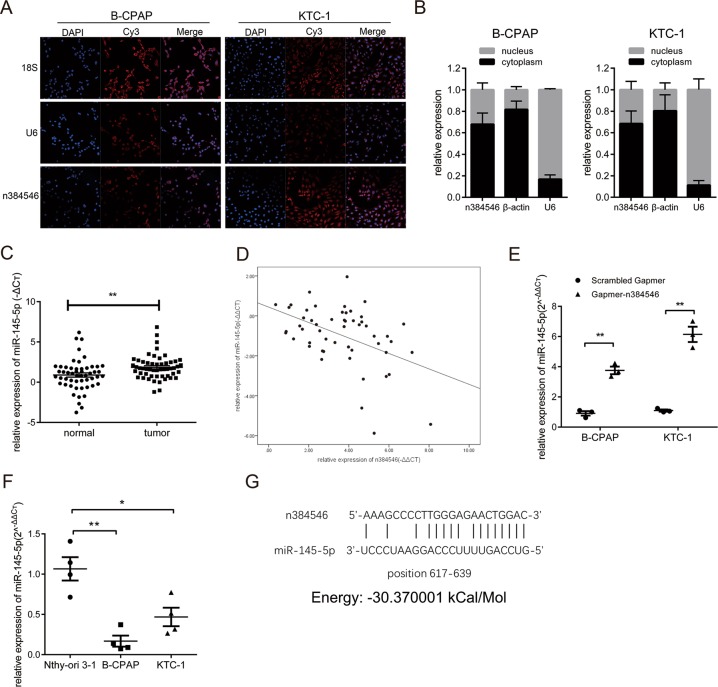
Fig. 4. MiR-145-5p is regulated by n384546.
a Localization of n384546 in B-CPAP and KTC-1 cells by fluorescent in situ hybridization. Nuclei are stained blue (DAPI) and n384546 is stained red. 18S is localized in the cytoplasm and U6 is localized in the nucleus. b The percentage of n384546, β-actin, and U6 in the cytoplasm and nucleus fraction of B-CPAP and KTC-1 cells was determined by qRT-PCR. c MiR-145-5p expression in 53 pair samples of PTC and adjacent normal tissues was determined by qRT-PCR. d Negative correlation between n384546 and miR-145-5p expression in PTC patients (Pearson Correlation Coefficient = −0.459, p < 0.01). e MiR-145-5p expression in Scrambled Gapmer or Gapmer-n384546 transfected B-CPAP and KTC-1 cells was determined by qRT-PCR. f Relative levels of miR-145-5p in normal thyroid cell Nthy-ori 3-1 and two types of PTC cells, B-CPAP and KTC-1 were determined by qRT-PCR. g The predicted binding sites and binding energy of miR-145-5p to the n384546 sequence. Data in (d) represent the mean ± SEM of three separate experiments. Data in (e) represent the mean ± SEM of four separate experiments. *p < 0.05, **p < 0.01 in paired Student’s t test (b) and independent Student’s t test (d, e)

