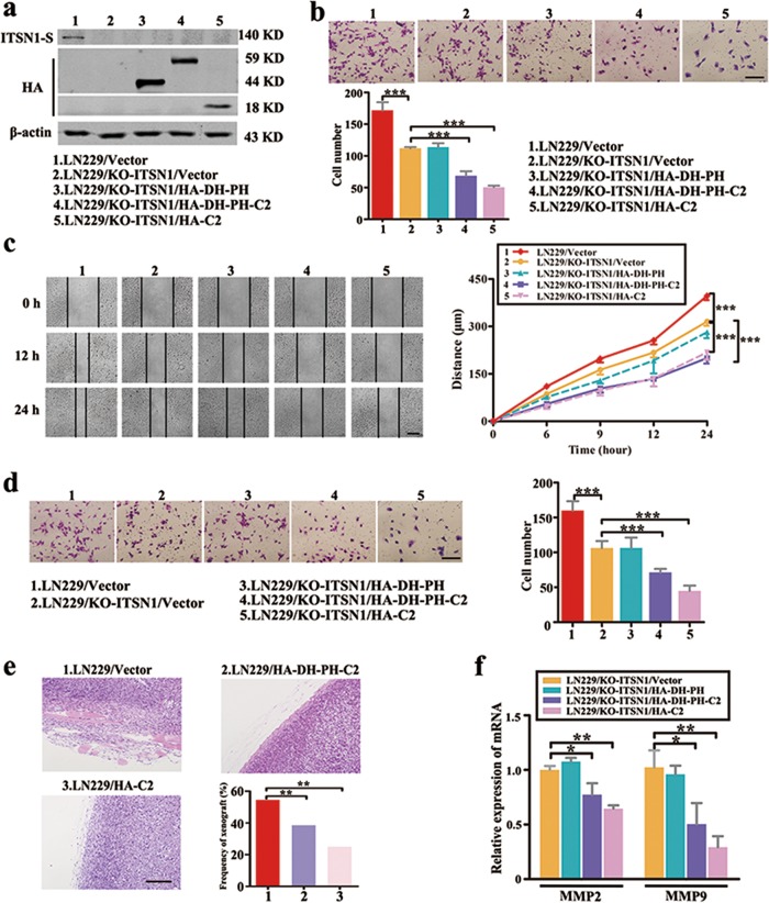
Fig. 3. Intersectin1 (ITSN1)-S and ITSN1-L displayed opposite roles in cell migration and invasion.
a Several exogenous different domain structure fragments of ITSN1-L were transfected into LN229/KO-ITSN1 cells and tested with anti-HA and anti-ITSN1-S antibodies in western blot. b Migration assay of the indicated cells. Cells migrating through transwell inserts were stained, photographed (×200), and quantified. c Results of scratch assay. The images were photographed at 0, 12, and 24 h (×100). d Invasion assay results. Cells invading through extracellular matrix-coated transwell inserts were stained, photographed (×200), and quantified. e Representative images of hematoxylin–eosin staining in LN229/Vector, LN229/HA-DH-PH-C2, and LN229/HA-C2 cells (×200). The frequency of xenograft invasion in mice was analyzed quantitatively. f Quantitative real-time PCR results of mRNA level of matrix metalloproteinase 2 (MMP2) and MMP9. GAPDH was used as control. Values were expressed as mean ± SD from three independent experiments (Student’s t test, *P < 0.05, **P < 0.01, ***P < 0.001). Scale bars, 200 μm

