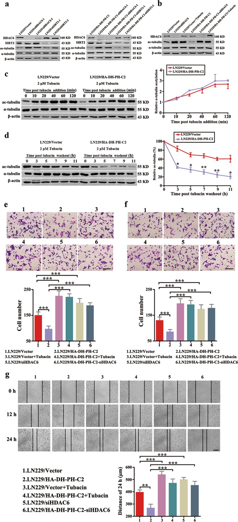
Fig. 5. C2 domain of intersectin1 (ITSN1)-L promoted microtubule deacetylation through activation of HDAC6.

a LN229 cells stably infected with lentivirus containing HDAC6 or SIRT2 shRNA sequences were lysed and analyzed by western blot. b Western blot of LN229 cells treated with shHDAC6 or 2 μM tubacin for 4 h. The level of HDAC6, ac-tubulin, and α-tubulin were examined. c Western blot (left panel) and quantitation (right panel) of the time course of α-tubulin acetylation after the addition of 2 μM tubacin. d Cells were treated with 2 μM tubacin for 12 h. Then tubacin was removed and the rate of α-tubulin deacetylation was tested by western blot (left panel) and quantitation (right panel). e–g Migration assay (e), invasion assay (f), and scratch assay (g) were performed in LN229 cells treated with shHDAC6 or 2 μM tubacin. Values were expressed as mean ± SD from three independent experiments (Student’s t test, *P < 0.05, **P < 0.01, ***P < 0.001). Scale bars, 200 μm
