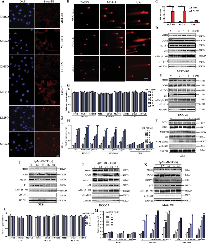
Fig. 4. Compound MI-743 causes intracellular 8-oxo-dG accumulation and DNA damage.
a MGC-803, HGC-27 and GES-1 cells were treated with DMSO or 5 µM MI-743 for 48 h. Intracellular 8-oxo-dG was stained with Cy3-conjugated avidin. DNA was counterstained with 4,6-diamidino-2-phenylindole (DAPI). Images were acquired at ×100 magnification by a Nikon Eclipse TE 2000-S fluorescence microscope. At least three independent experiments were performed for each group. b MGC-803, HGC-27 and GES-1 cells were treated with DMSO or 10 µM MI-743 for 48 h and run in alkaline comet assay. Pictures were originally captured at ×40 magnification. H2O2 was used as positive control. c Tail moment was calculated by CometScore software. Three individual experiments were performed for each group. d–f Western blot analysis of the protein levels of MTH1, MUTYH, OGG1, p21, ATMpS1981 and p53pS15 in MGC-803, HGC-27 and GES-1 cells, treated with increasing concentrations of MI-743 (0, 1, 2, 4, 8 and 12 µM). g, h Densitometry shows relative protein expression normalized for GAPDH. Data are representative of three independent experiments. i–k Western Blot analysis of the protein levels of MTH1, MUTYH, OGG1, p21, ATMpS1981 and p53pS15 of protein lysates, isolated from MGC-803, HGC-27 and GES-1 cells, which were treated with 12 µM MI-743 for 0, 12, 24, 36 and 48 h. l, m Densitometry shows relative protein expression normalized for GAPDH. Data are presented as means ± SD. Three individual experiments were performed for each group. *P < 0.05, **P < 0.01, ***P < 0.001 as compared with the controls

