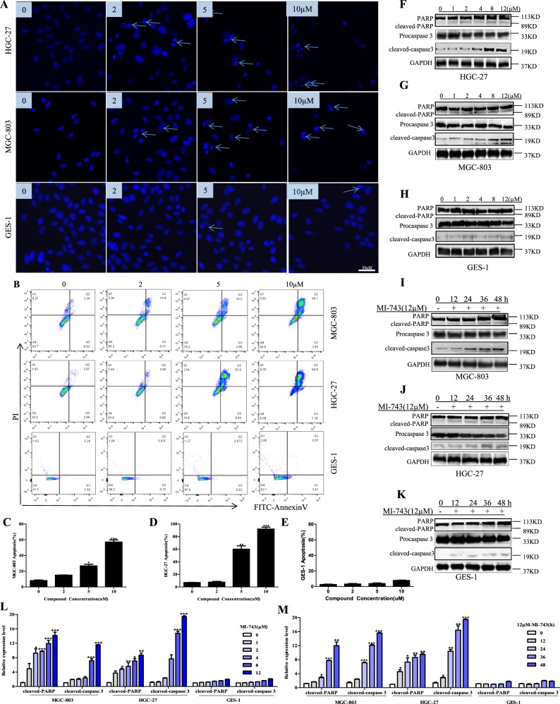
Fig. 5. Compound MI-743 induces MGC-803 and HGC-27 cell apoptosis.
MGC-803, HGC-27 and GES-1 cells were cultured with DMSO, 2, 5, 10 μM of MI-743 for 48 h. a Apoptosis-related nuclear condensation were stained with Hoechst 33258 and pictured by fluorescence microscopes. Pictures were originally captured at ×100 magnification. Representative photographs of three independent experiments were shown. b–e Apoptotic cells were detected using the Annexin V-FITC/ PI double staining and analyzed by flow cytometry in MGC-803, HGC-27 and GES-1 cells. The data were analyzed by FlowJo-V10 and Graph Pad Prism 5 software. Three individual experiments were performed for each group. f–h Western blot analysis of the protein levels of pro and cleaved-caspase 3, PARP and cleaved-PARP in MGC-803, HGC-27 and GES-1 cells, treated with increased concentrations of MI-743 (0, 1, 2, 4, 8 and 12 µM). l Densitometry shows relative protein expression normalized for GAPDH. Data are representative of three independent experiments. i–k Western Blot analysis of the protein levels of pro and cleaved-caspase 3, PARP and cleaved-PARP of protein lysates, isolated from MGC-803, HGC-27 and GES-1 cells, which were treated with 12 µM MI-743 for 0, 12, 24, 48 and 72 h. m Densitometry shows relative protein expression normalized for GAPDH. The data are presented as means ± SD. Three individual experiments were performed for each group. *P < 0.05, **P < 0.01, ***P < 0.001 as compared with the controls

