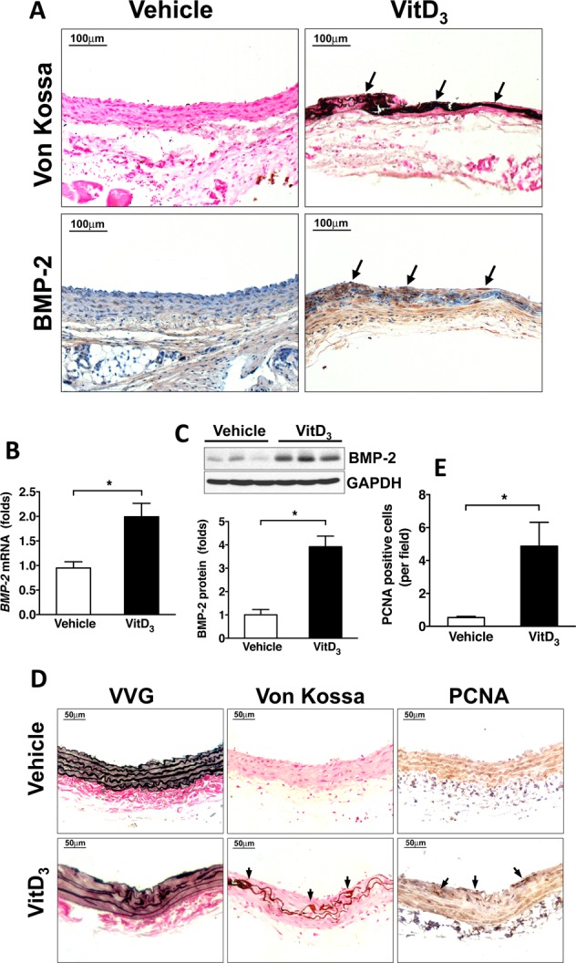
Figure 5.

BMP-2 expression is increased in calcifying carotid arteries from vitamin D3-injected rats. Male SD rats were injected with vehicle or 5 mg/kg vitamin D3 (VitD3) for 3 consecutive days, and carotid arteries were harvested 14 days later. (A) Immunohistochemical staining showing increased BMP-2 expression in calcifying areas after vitamin D3 injection. (B) qPCR results showing increased BMP-2 mRNA levels in vitamin D3 injected rats. (C) Western blotting results showed that vitamin D3 injection increased BMP-2 protein levels. (D) Vitamin D3 injection increased cell proliferation in calcifying areas but did not cause neointimal hyperplasia. Neointimal hyperplasia was evaluated by vessel wall thickness. Elastin integrity was examined by VVG staining. Calcification was assessed by Von Kossa staining. Cell proliferation was determined by PCNA immunohistochemical staining. (E) Quantitative data showing increased PCNA-positive cells in vitamin D3-injected rats.
