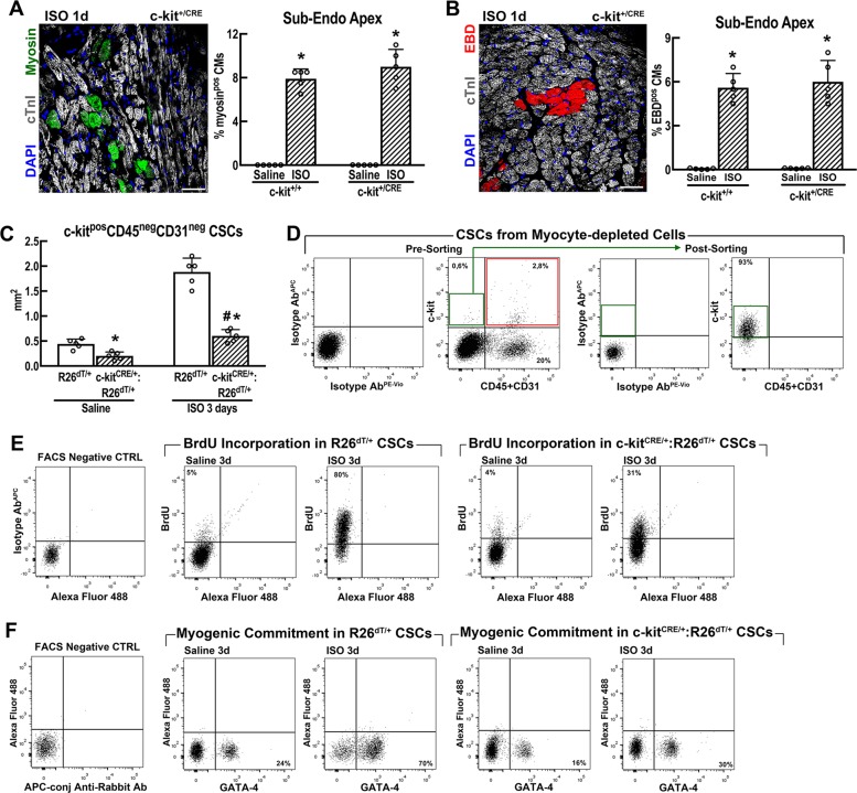
Fig. 5. c-kitCre mice show a defective CSC activation after injury in vivo.
a, b Necrotic cardiomyocytes (CM) (revealed either by myosin Ab in vivo labelling in a and EBD incorporation in b) were similarly increased in wild-type c-kit+/+ and heterozygous c-kit+/Cre mice at 1 day after ISO (200) in the Apex sub-endocardium. n = 5 mice per group, *p < 0.05 vs. saline (one-way ANOVA analysis with Tukey’s multiple comparison test). Scale bars = 50 µm. c c-kitCreGFPnls:R26floxed-dTomato/+ (abbreviated as c-kitCre/+:R26dT/+) mice show a blunt increase of resident CD45negc-kitpos CSCs when compared with control R26floxed-dTomato/+ (abbreviated hereafter as R26dT/+) mice 3 days after ISO. n = 5 mice per group; *p < 0.05 vs. Saline; #p < 0.05 vs. R26dT/+ mice (one-way ANOVA analysis with Tukey’s multiple comparison test). d Representative FACS detection and isolation of CSCs from cardiomyocyte-depleted cardiac cell preparations. Left, representative gating strategy using isotype antibodies. Pre-sorting, flow cytometry dot plots show that the majority of total c-kitpos cardiac cells are CD45 or CD31 positive (red box), whereas only a minority are CD45 and CD31 negative (green box). Post sorting, flow cytometry dot plots show that CD45posCD31pos (linpos) cells are efficiently removed from cardiac cells by CD45−CD31 sorting and CD45negCD31negc-kitpos-sorted cells uniformly express low levels of c-kit. e Flow cytometry dot plots (representative of n = 3) show BrdU incorporation in CD45neg c-kitpos cardiac cells from saline and ISO-treated R26dT/+ and c-kitCre/+:R26dT/+mice. BrdU—35mg/Kg bid—was intraperitoneally administered in vivo for 3 days in adult mice every 12 h before killing. f Flow cytometry dot plots (representative of n = 3) show the fraction of myogenic-committed Gata-4pos CD45negc-kitpos CSCs isolated from saline and ISO-treated R26dT/+ and c-kitCre/+:R26dT/+ mice. All data are mean ± SD

