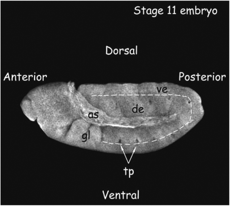
Fig. 2.
Stage 11 embryo. This embryo was stained with Texas Red–conjugated Wheat Germ Agglutinin (Molecular Probes) to visualize major morphological markers. The dorsal and ventral epidermis (de and ve, respectively) are marked. The gnathal lobes (gl) and trachael pits (tp) are characteristic of this stage embryo. as = amnioserosa. Embryo staging is as in (13).

