Fig. 2.
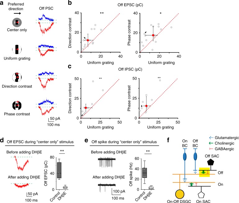
Contextually sensitive cholinergic excitation underlying the differential modulation of pDSGC Off response. a Whole-cell recording traces of Off IPSCs (blue) and Off EPSCs (red) from a pDSGC in a control mouse. PSC traces represent trial average (darker traces) and SEM (lighter traces) for this and subsequent figures. b Scatter plots compare Off EPSC charge transfer between uniform grating and compound gratings. Direction-contrast vs uniform grating: n = 15 cells from 11 mice, **p = 0.0043; Phase-contrast vs uniform grating: n = 15 cells from 9 mice, *p = 0.035 (Wilcoxon signed-rank test). c Same as b, but for Off IPSC charge transfer. Direction-contrast vs uniform grating: n = 9 cells from 5 mice, **p = 0.0039; Phase-contrast vs uniform grating: n = 10 cells from 6 mice, **p = 0.0098. d Left: Example Off EPSC traces of a pDSGC during center only grating before and after adding DHβE. Right: Summary graph of pDSGC Off excitatory charge transfer in control (Ames’ solution, n = 4 cells from 2 mice) or in the presence of DHβE (n = 7 cells from 2 mice): **p = 0.0061 (Wilcoxon rank-sum test). See Supplementary Fig. 2a for Off IPSC as a negative control. e Left: Example Off spike traces of a pDSGC during center only grating before and after adding DHβE. Right: Summary graph of pDSGC Off spike firing rate before (in Ames’ solution) or after adding DHβE: n = 10 cells from 6 mice, **p = 0.0020 (Wilcoxon signed-rank test). f Schematic shows the synaptic connections modulated by motion context in the Off pathway (yellow filled rectangle). Glutamatergic BC inputs are shown in dotted arrows indicating their contribution is little under the experimental conditions in this study. See also Supplementary Fig. 2c for Off EPSC charge transfer under different stimulus contexts in the present of DHβE

