Figure 5.
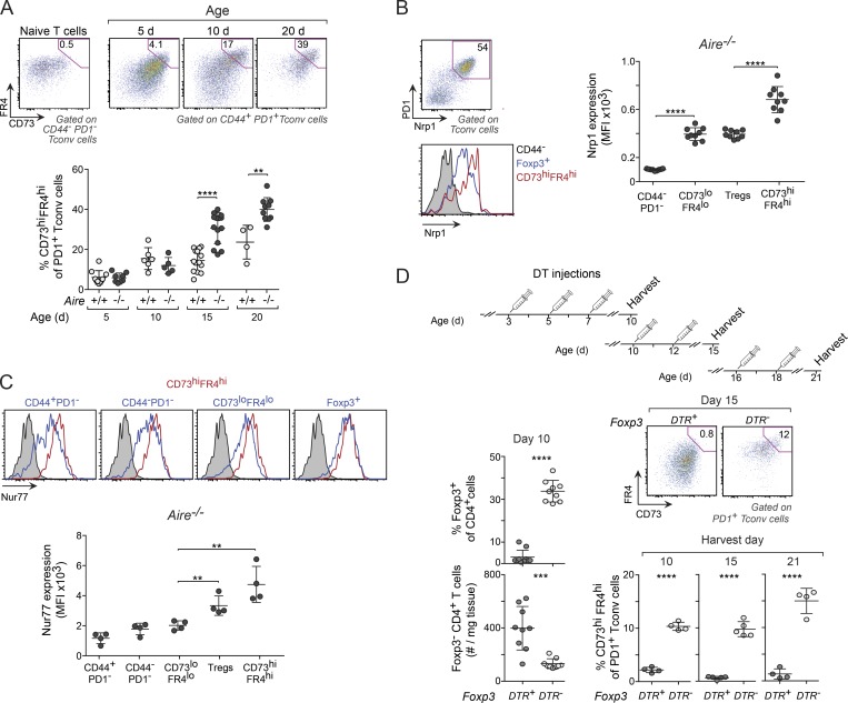
Perinatal T reg cells were critical for maintaining anergy in PD-1+ T conv cells. (A) Frequencies of anergic (CD73hiFR4hi) PD-1+ T conv cells in liver from mice of various ages. Top: Representative flow-cytometric plots showing gating strategy using CD44−PD-1− T conv cells to identify anergic cells. Bottom: Summary data (n = 4–14 mice/group). (B) Nrp1 expression on various populations of liver CD4+ T cells from 10-d-old Aire−/− mice (n = 9 mice/group). (C) Expression of Nur77 in cells from 10-d-old Nur77eGFP reporter mice (n = 4 mice/group). Nur77eGFP− cells were used as controls in all histograms (gray shadings). MFI, mean fluorescence intensity. (D) Frequency of anergic T conv cells in T reg cell–depleted perinates. Top: Treatment regimen for DT injection. Bottom left: T conv and T reg cell summary data on DT-treated 10-d-old mice. Right: Representative flow-cytometric plots and summary data for anergic T conv cells from DT-treated mice (n = 4–9 mice/group). Data are pooled from two to four independent experiments and show mean ± SD. Statistical analyses as in Fig. 1. **, P ≤ 0.01; ***, P ≤ 0.001; ****, P ≤ 0.0001.

