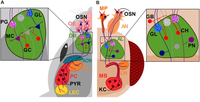
Figure 1.
Diagram of the mouse and Drosophila olfactory systems. Sensory neurons (OSN) in the nose or antennas (AN) and maxillary palp (MP) project to the superior centers of olfactory sensory processing, olfactory bulb (OB) and antennal lobe (AL) in mice (A) and Drosophila (B), respectively. Mitral cells (MC) send their axons directly to the accessory olfactory nucleus (AON), olfactory tubercle (OT), pyriform cortex (PC) and lateral entorhinal cortex (LEC), while projection neurons (PN) project to the mushroom body (MB) and lateral horns (LH). Olfactory epithelium (OE); glomerulus layer (GL); periglomerular cell (PG); granule cell (GC); GABAergic neurons (GB); cholinergic neurons (CH); kenyon cells (KCs). Neurons that are synaptically connected are depicted in the same colors.

