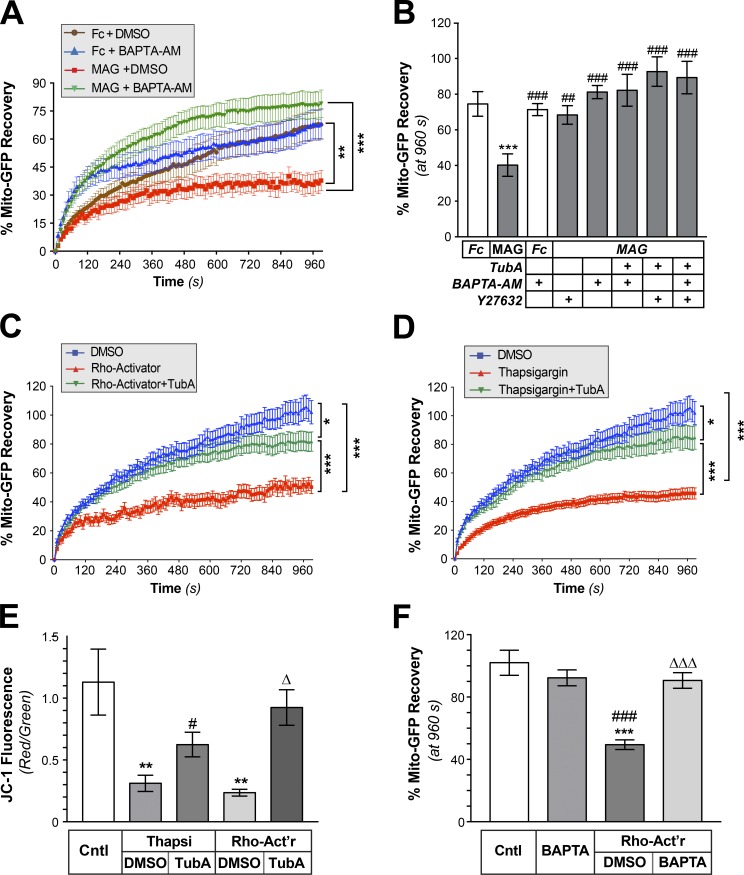
Figure 5.
RhoA/ROCK pathway activates HDAC6 through a Ca2+-dependent mechanism. (A) FRAP analyses for Mito-GFP in distal axons of DRGs cultured on laminin and treated with bath-applied Fc + DMSO, Fc + 3 µM BAPTA-AM, MAG-Fc + DMSO, or MAG-Fc + 3 µM BAPTA-AM are shown as average normalized percentage recovery ± SEM (n ≥ 9 axons over three culture preparations; **, P ≤ 0.01; ***, P ≤ 0.005 for indicated treatments by two-way ANOVA with Tukey post hoc). BAPTA-AM is not statistically different from over 300 to 960 s. (B) End-point FRAP for Mito-GFP in distal axons of DRGs treated with bath-applied Fc vs. MAG-Fc ± 3 µM BAPTA-AM, 10 µM Y27632, 10 µM TubA, or indicated combinations of these inhibitors is shown as average of normalized percentage recovery ± SEM at 960 s after bleach (n ≥ 9 axons over three culture preparations; ***, P ≤ 0.005 vs. Fc; ##, P ≤ 0.01; ###, P ≤ 0.005 vs. Mag-treated by two-way ANOVA with Tukey post hoc). BAPTA-AM is not statistically different from vehicle. (C and D) FRAP for Mito-GFP in distal axons of DRGs treated with RhoA Activator (C) or Thapsigargin (D) is shown as average normalized percentage recovery ± SEM. Data for vehicle control (DMSO), 1 µg/ml of Rho-Activator (+ DMSO), Rho-Activator + 10 µM TubA, 1 µM Thapsigargin (+ DMSO), and Thapsigargin + 10 µM TubA are shown (n ≥ 9 axons over three culture preparations; *, P ≤ 0.05; ***, P ≤ 0.005 for indicated treatments by two-way ANOVA with Tukey post hoc). (E) Quantification of mitochondrial membrane potential based on red/green fluorescence of JC-1 in axon shafts of DRGs cultured on laminin (Cntl) or aggrecan (CSPG) substrates is shown after treatment with 1 µM Thapsigargin (Thapsi), Thapsi + 10 µM TubA, 1 µg/ml Rho-Activator (Rho-Act’r), or Rho-Act’r + TubA. Values represent average ratio of normalized red/green fluorescent JC-1 signals ± SEM (n ≥ 20 axons over three culture preparations; **, P ≤ 0.01 vs. control; #, P ≤ 0.05 vs. Thapsi + DMSO; Δ, P ≤ 0.01 vs. Rho-Act’r + DMSO by two-way ANOVA with Tukey post hoc). (F) End-point FRAP for Mito-GFP in distal axons of DRGs cultured on laminin and treated ± 3 µM BAPTA-AM, 1 µg/ml Rho-Act’r, Rho-Act’r + BAPTA-AM is shown as average of normalized percentage recovery ± SEM at 960 s after bleach (n ≥ 9 axons over three culture preparations; ***, P ≤ 0.005 vs. control; ###, P ≤ 0.005 vs. BAPTA-AM; ΔΔΔ, P ≤ 0.005 vs. Rho-Act’r + DMSO by two-way ANOVA with Tukey post hoc). BAPTA-AM is not statistically different from control.

