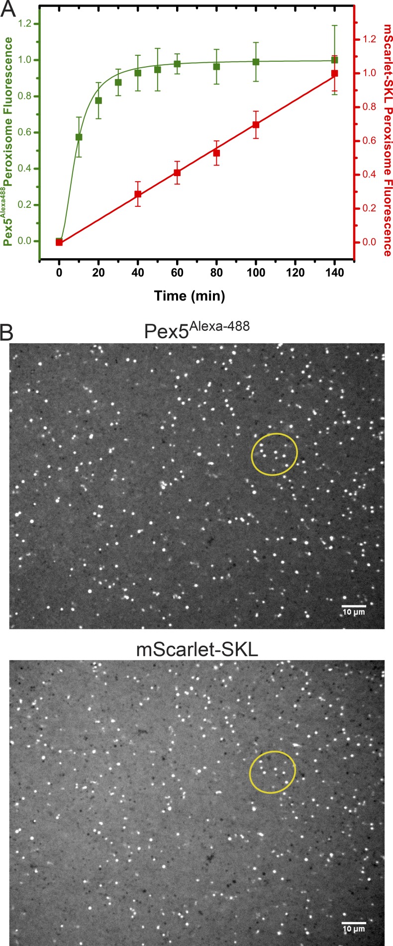
Figure 5.
Kinetics of Pex5 binding and peroxisome protein import. (A) mScarlet-SKL (0.6 µM) and fluorescently labeled Pex5 (Pex5Alexa488; 0.15 µM) were added at time point zero to cleared Xenopus egg extract. The sample was mounted on a PEG-passivated glass chamber and imaged over time using a spinning-disk confocal microscope. The red (mScarlet-SKL) and green (Pex5Alexa-488) emission channels were imaged simultaneously. Shown are the combined data of two different experiments (>20 images per time point), each normalized to the final time point of the control. For each time point, the mean and the standard deviation of the mean are given. (B) Images taken at 140 min sequentially in the two channels. The foci partially overlap (compare foci inside the oval). Overlap is not perfect because the peroxisomes are moving. Bars, 10 µm.

