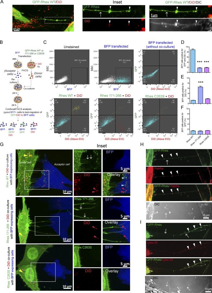
Figure 5.
Rhes-induced TNT-like Rhes tunnels contain DiD-, Rab5a-, or Lyso-20–positive vesicles. (A) Representative confocal image of striatal neuronal cells expresing GFP-Rhes and treated with DiD-Red (DiD, vesicle labeling dye). Arrows point to TNT-like protrusions, and arrowheads point to vesicles in TNT-like cellular protrusions. (B) Experimental design for C. (C) FACS analysis of striatal neuronal cells expressing GFP-Rhes WT, GFP-Rhes 171–266, or GFP-Rhes C263S treated with DiD and co-cultured with FACS-sorted BFP-transfected striatal neuronal cells. The BFP population was gated and analyzed for the presence of GFP and/or DiD (Alexa Fluor 633). (D–F) Bar graphs show data mean ± SEM; one-way ANOVA test (***, P < 0.001), n = 3. The quantification of BFP cells positive for both GFP and DiD fluorescence (D), only DiD (E), or only GFP (F). (G) Representative confocal images for co-cultured experiments as indicated in B (see Results). (H and I) Representative confocal and DIC images of striatal neuronal cells cotransfected with GFP-Rhes and Rab5a (H) or GFP-Rhes and Lyso-20 (I). Arrows indicate TNT-like Rhes tunnels, and arrowheads show vesicles in Rhes tunnels.

