Figure 8.
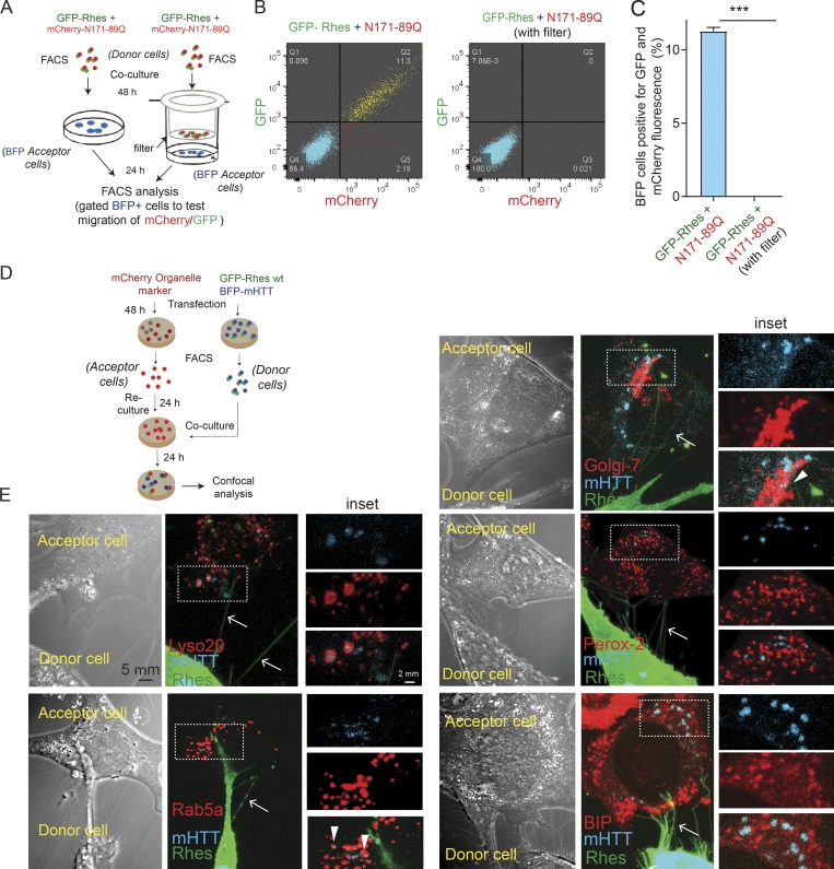
Rhes requires physical cell–cell contact to transport mHTT, which in acceptor cells associates with lysosome and other vesicles. (A) Experimental design for B and C. (B) FACS plot for the indicated co-cultured striatal neuronal cells. BFP cell population was gated, and 20,000 cells were recorded per sample. (C) Bar graphs show data mean ± SEM; Student’s t test (***, P < 0.001), n = 3, indicating percentage of BFP cells (acceptor cells) positive for Rhes + N171 89Q and Rhes + N171-89Q with Transwell (filter). (D) Experimental design for E. (E) Representative confocal and DIC images and their insets indicating donor cell (GPF-Rhes/BFP-mHTT) and acceptor cell (mCherry-tagged various organelle markers). Green channel shows GFP-Rhes, blue color represents BFP-mHTT, and red channel indicates the mCherry-tagged Lysosome (Lyso 20), Rab5a, Golgi-7, Peroxisome (Perox-2), and BIP individual organelle markers. Arrows indicate colocalization between BFP-mHTT and different organelle markers.

