Figure 4.
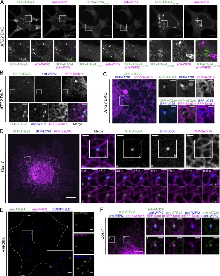
Overexpressed GFP-ATG2A and endogenous ATG2A localizes at ER–autophagosomal membrane contact sites. (A) ATG2 DKO HEK293 cells stably expressing the indicated GFP-ATG2A construct were treated with OA to enlarge LDs, and then incubated in starvation medium (Earle’s balanced salt solution) and subjected to anti-WIPI2 IF. Greater than 88% of all WIPI2 positive structures are also GFP-ATG2A positive. Targeting to autophagosomes (WIPI2 structures) is lost with removal of the mini-ATG2 sequence (GFP-ATG2AΔ1–345). Scale bars: 10 µm; zoomed field: 1 µm. (B) GFP-ATG2A localized with early autophagosome markers is also associated with the ER. Anti-WIPI2 IF confocal images in starved ATG2 DKO cells stably expressing GFP-ATG2A and transiently expressing RFP-Sec61b. Scale bars: 1 µm. (C) The late autophagosome marker LC3B colocalized with GFP-ATG2A ∼13% of the time, and these LC3B/GFP-ATG2-positive puncta consistently also associated with the ER. Live imaging of starved ATG2 DKO cells stably expressing GFP-ATG2A, transiently expressing RFP-Sec61b and BFP-LC3B, and incubated with OA. Note that ring-like GFP-ATG2 structures are likely LDs, are not LC3B-positive, and are not restricted to the ER. Full time course in Fig. S3 B indicates GFP-ATG2A remains directly associated with the ER during the entire time it localizes to the autophagosome. Scale bars: 1 µm. (D) Time-lapse imaging of starved Cos-7 cells stably expressing GFP-ATG2A and transiently expressing RFP-Sec61b and BFP-LC3B. ER localization of autophagosome-associated GFP-ATG2A is similar in WT Cos-7 cells as in DKO HEK293 cells, and again persists throughout the residence time of GFP-ATG2A at LC3B-positive structures. A second longer video example is shown in Fig. S3 C. Scale bars: 10 µm; zoomed field: 1 µm. (E) Endogenous ATG2A is localized only on autophagosomes (WIPI2-positive) and not LDs. Commercially available ATG2A antibodies were precleared with ATG2 DKO cellular material to improve signal to noise (Fig. S3). Then, starved HEK293 cells were treated with OA, labeled with BODIPY 558/568, and subjected to anti-WIPI2 and precleared anti-ATG2A IF. Scale bars: 10 µm; zoomed field: 1 µm. (F) Endogenous ATG2A is associated with autophagosome markers and the ER. Starved Cos-7 cells were transiently transfected with RFP-Sec61b, treated with OA, and subjected to anti-WIPI2 and precleared anti-ATG2A IF. Scale bars: 5 µm; zoomed field: 1 µm. For all panels, representative confocal images from at least three independent experiments are shown.

