Figure 1.
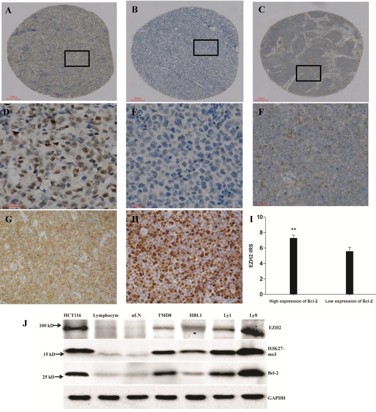
Expression of EZH2, Bcl-2 and Ki-67 in diffuse large B-cell lymphoma (DLBCL) and normal lymph node (nLN) tissues, scale bar=200 μm for A-C and scale bar=20 μm for D-H. (A). High expression of EZH2 in DLBCL with more than 90% tumor cells intensively stained by EZH2 antibody in nuclei; (B). Negative immunostaining of EZH2 in DLBCL; (C). No EZH2 staining in nLN. (D-F). High-power images of the areas demarcated in boxes in A-C. (G-H). Positive immunostaining of Bcl-2 and Ki-67 in DLBCL tissues. Bcl-2 expression exhibited a cytoplasmic pattern and Ki-67 showed a distinct nuclear pattern. (I) Correlation between expression of EZH2 and Bcl-2 in DLBCL. The staining score of EZH2 was significantly higher in high Bcl-2 expression patients compared to those of low Bcl-2 expression (7.29±0.36 and 5.58±0.50, respectively, P=0.005). (J) EZH2, H3K27me3 and Bcl-2 protein expression was detected by western blot in colon cancer cell line HCT116, human peripheral blood lymphocyte, normal lymph node (nLN) and 4 DLBCL cell lines (TMD8, HBL-1, Ly1 and Ly8). Lysates from the positive control HCT116 (lane 1), and tumor cell lines (lanes 4-7) had much higher EZH2, H3K27me3 and Bcl-2 immunoreactivity than normal human peripheral blood lymphocyte (lane 2) and nLN tissue (lane 3).

