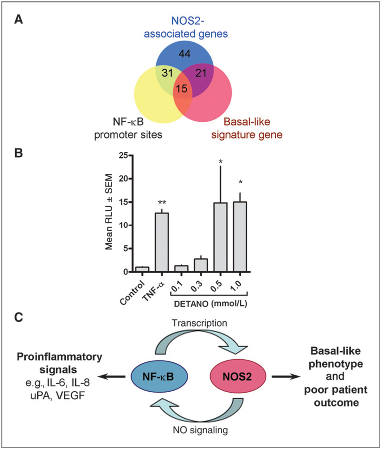
Figure 1.
Nitric oxide stimulates NF-κB activity in ER− breast cancer cells. A, Venn diagram depicting the relationship between genes overexpressed in high NOS2 tumors, promoter NF-κB sites, and the expression of basal-like signature genes. B, MDA-MB-468 cells were transiently transfected with an NF-κB luciferase reporter construct and exposed to TNF-α (20 ng/mL) or DETANO for 24 hours. Luciferase activity shown as mean RLU ± SD was normalized to untreated control and the fold increase relative to control is presented. Statistical significance (*, P < 0.05) compared with control was determined by one-way ANOVA. C, scheme representing the positive feedback loop between NOS2 and NF-κB activity and the products of this reciprocal signaling on tumor inflammation and patient outcomes.

