Figure 2.
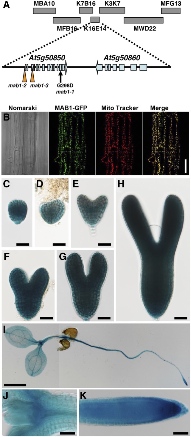
MAB1 encodes a mitochondrial PDC E1β. A, Map position of the MAB1 locus. MAB1 was mapped between the MBA10 and MFG13 bacterial artificial chromosomes on chromosome 5 by map-based cloning. mab1-1 is a point mutation in At5g50850 that causes the amino acid change G298D. In mab1-2 and mab1-3 mutants, T-DNA is inserted ∼310 bp upstream of the translation start site and at the third exon of the gene, respectively. B, Localization of MAB1-GFP and Mito Tracker fluorescence in the epidermis of Arabidopsis roots. The Nomarski image (left), and GFP (second from left) and Mito Tracker (second from right) fluorescence images were obtained using an epifluorescence microscope. At right is a merge of the MAB1-GFP and Mito Tracker images. C to K, Expression patterns of MAB1p:GUS in the embryo at the globular (C), transition (D), heart (E), late heart (F), and torpedo (G, H) stages, and in the 4-d-old seedling (I), around shoot apical meristem (J) and root meristem (K), respectively. GUS staining was performed at 37°C overnight (C to H) and for one hours (I to K). Scale bars = 50 mm (B, J, K), 1 cm (I), and 20 mm (C to H).

