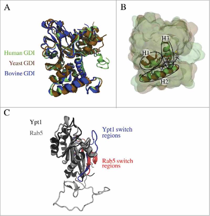
Figure 2.

(A) Superposition of modelled human GDI (green) and X-ray structures of yeast GDI (PDB entry 2BCG,17 brown) and bovine GDI (PDB entry 1LV0,15 blue). (B) The geranylgeranyl binding pocket is formed by three helices (H1, H2, and H3) and accommodates the prenyl moieties of the small GTPase (from yeast 2BCG in brown, the modelled geranylgeranyl chains are coloured according to the atom types). (C) Superposition of Rab5 (grey) and Ypt1 (black) revealed structural differences in the GTPase switch I region coloured in red (Rab5) and blue (Ypt1), respectively. This is due to the fact that for Rab5(GDP) in complex with Rabaptin5 a conformation with an unusual β-strand in the switch I region was crystallized (PDB entry 1TU422).
