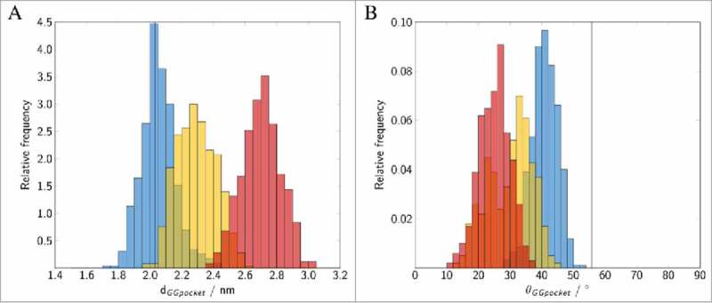
Figure 8.

Binding of geranylgeranylated Rab5 to the GG-binding pocket of GDI. Distribution of (A) the distance between the GG chains and Met132, (B) the angle θ between helices H1 and H2 forming the GG-binding pocket. The vertical lines correspond to the GG-binding pocket distance and angle observed in the yeast complex from 2BCG.17 Data for the individual runs are coloured in blue (cytRun1), yellow (cytRun2) and red (cytRun3).
