Figure 13.

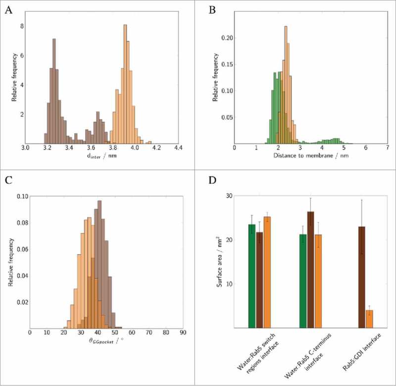
Distribution of (A) intermolecular distances of cytoplasmic (brown) and membrane-bound Rab5(GDP):GDI complex (orange). (B) Distances between the membrane and un-complexed Rab5(GDP) (green) and Rab5(GDP):GDI (orange). (C) Distribution of the opening angle θ of the GG-binding pocket in cytoplasmic (brown) and membrane-bound complex (orange). (D) The solvent accessible surface area (SASA) of the Rab5 switch regions and HVR as well as the total Rab5(GDP):GDI protein-protein interface area averaged over 200 ns of MD simulations. Data for un-complexed Rab5 are shown in green, for the cytoplasmic tightly bound Rab5(GDP):GDI complex in brown, and for the membrane-bound complex in orange. Data were averaged over three 500 ns MD simulations for un-complexed Rab5 from previous studies.
