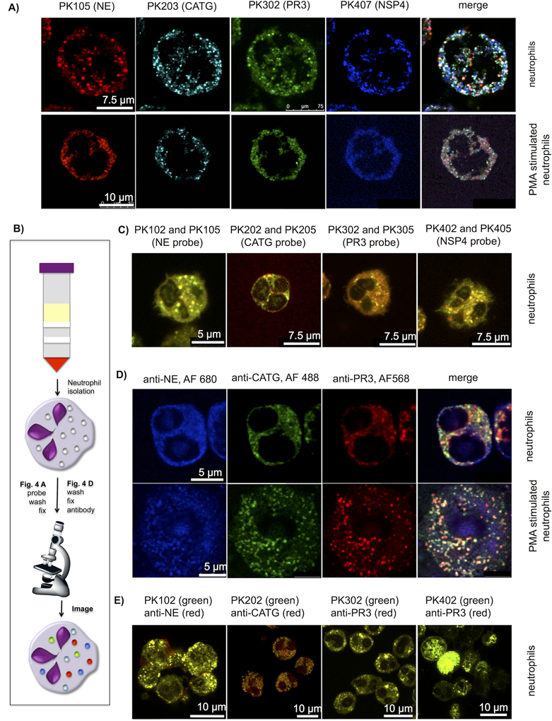
Figure 4. Localization of active NSPs in neutrophil granules.
(a) Freshly isolated neutrophils were allowed to settle on cover slips, treated or untreated with PMA for 3 hours, incubated with the indicated probes (50nM) for 30 min and fixed (b) scheme of experimental flow (c) Neutrophils labeled with two probes (100nM each) with the same sequence and two different fluorescent dyes and fixed. Each signal overlays well, demonstrating specific probe labeling of the targeted enzyme. For separate channels see Supporting Information Figure S6. (d) Neutrophil staining with antibodies for three individual NSPs. Neutrophils were prepared as in (a) without probe labeling, stained with anti-NE, anti-PR3 and anti-CATG followed by secondary antibodies with the fluorophores indicated in the figure. (e) Co-staining with activity-based probes and antibodies. Neutrophils were allowed to settle on cover slips, treated with 50nM of the indicated probes, fixed and stained with anti-NE, anti-PR3, anti-CATG and anti-NSP4 respectively, followed by a secondary antibodies with the indicated fluorophores. (For separate channels see Supporting Information Figure S7). All slides were mounted and imaged by confocal microscopy with 488nm, 552nm and 648nm lasers. Images are representative of 3 separate donors (For more examples and z-stack movies see Supporting Information Figure S5–S7 and Supporting Information movies S1–S4)

