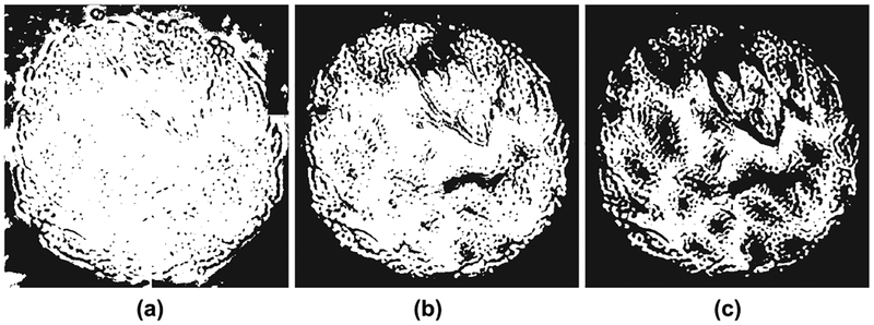
Fig. 3:
Background subtraction for various spectral energy thresholds: (a) 0.1, (b) 0.35, and (c) 0.7. A small threshold counts some background pixels as tissue (white), and a large threshold marks some tissue pixels as background (black). Compare to Figure 1.

