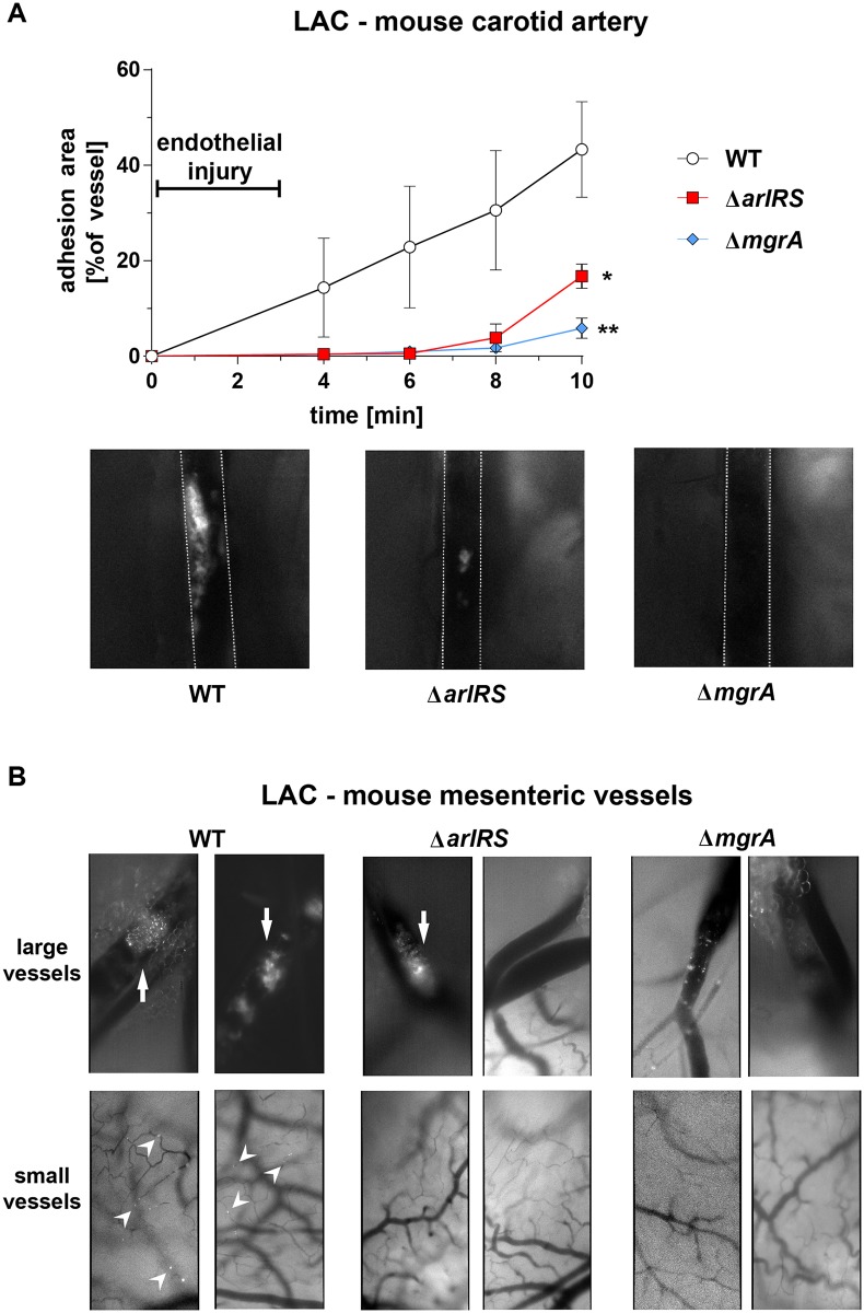
Fig 8. ArlRS–MgrA cascade controls adhesion to vasculature in vivo.
Adhesion of GFP-expressing S. aureus LAC WT and its mutants to mouse carotid artery (A) with FeCl3-injured endothelium was monitored using intravital microscopy after intravenous injection of fluorescent bacteria. Representative microscopy images of artery at 8 min time-point are shown (image size: 2 mm × 2 mm; outlines of arteries marked with white lines). N = 3 per group. *p<0.05, **p<0.01, compared to WT. Data presented as mean ± SEM. Adhesion of GFP-expressing S. aureus LAC WT and its mutants to a Ca2+-ionophore-activated mouse mesenteric circulation (B) was evaluated using intravital microscopy after intravenous injection of fluorescent bacteria. Two independent experiments (2–3 mice per group) were performed, and representative images at 30 min post-injection are shown (image size: 0.5 mm × 1 mm). Large adherent clots were observed in large mesenteric vessels after injection of WT and ΔarlRS bacteria (arrows). Small clumps and individual adherence of WT bacteria was visible in small vasculature (arrowheads). No adherence of ΔarlRS and ΔmgrA mutants in small vessels was observed.

