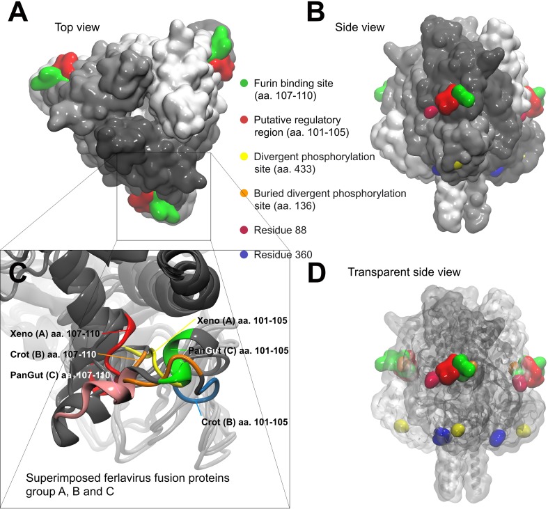
Fig 3. Ferlavirus fusion protein trimer models.
The furin binding site (green) and the newly described putative regulatory region (red) located at the edges of the triangle shaped fusion protein trimer complex. The divergent predicted phosphorylation sites are also indicated on the models. These sites can be seen from top (A) and side views (B) and a transparent side view (D) was rendered to see the buried divergent phosphorylation site. Variations of two residues (88 and 360) significantly alter the electrostatic charge distribution around the furin binding site. Panel C shows the structural differences between the three studied ferlavirus groups around the furin binding site.

