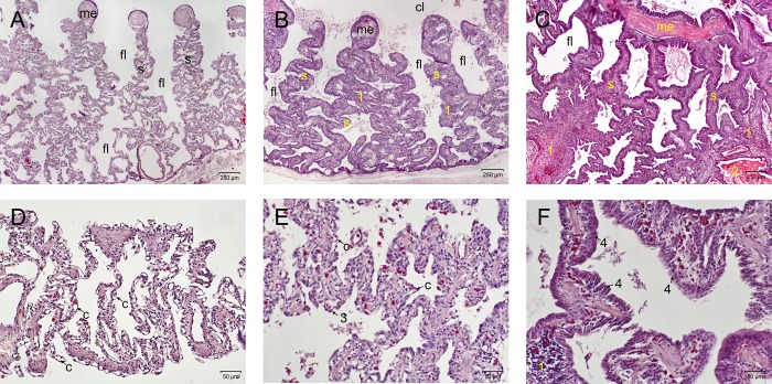
Fig 6.
Sections of the lung, light microscopy, H&E stain, A-C: 40x magnification; D-F: 200x magnification, day post infectionem. (A,D) Score 0, snake No. P1, day 49. Demonstration of a cross section through an unremarkable lung. The myoelastic inner border of the lung tissue and faveoli lead to the respiratory tissue along the septae. Capillaries are bulging into the air spaces, demonstrating a thin air-blood-barrier. (B,E) Score 1, snake No. A4, day 16. Demonstration of a cross section through a ferlavirus infected lung, moderate changes. The septae appear thickened and initial transformation of the lung epithelium can be seen. Capillaries are still close to the surface, but the epithelial cells are starting to transform (3). Infiltration of lymphocytes and plasma cells in the septae (1). (C,F) Score 2, snake No. B11, day 35. Demonstration of a cross section (not complete) through a severely affected lung. The septae are massively thickened, the faveoli narrowed and partially filled with debris (2). Massive thickening and infiltration of heterophils, lymphocytes and plasma cells in the interstitium (1), transformation of the respiratory epithelium into a multilayered epithelium (4). (c–capillary; cl–central lumen; fl–faveolus; me—myoelastic tissue; s—septum).

