Figure 5.

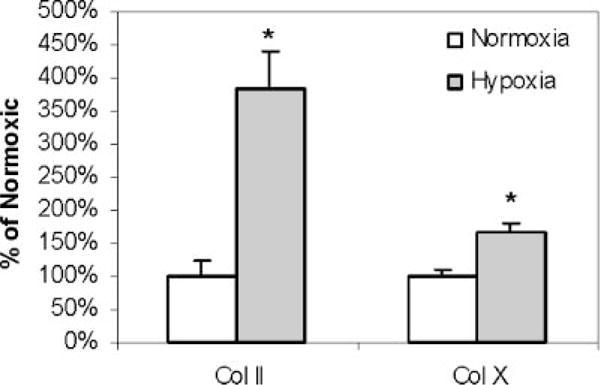
Quantification of type II and type X collagen immunofluorescence. Means and SD for quantitative integration of immunofluorescence in seven beads for each condition are shown. *p < 0.05 versus normoxia.

Quantification of type II and type X collagen immunofluorescence. Means and SD for quantitative integration of immunofluorescence in seven beads for each condition are shown. *p < 0.05 versus normoxia.