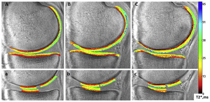
Fig. 3.
Sample mid-sagittal ultrashort echo time (UTE)-T2* maps of an uninjured control subject (A,a), the contralateral uninjured knee of an anterior cruciate ligament reconstruction (ACLR) subject (B,b), and the anterior cruciate ligament (ACL)-reconstructed knee of the same ACLR subject (C,c). Regions of interest employed in UTE-T2* profile analyses from the ACLR subject’s contralateral (b) and ACL-reconstructed (c) knees demonstrate irregularities from the relatively smooth laminar distribution of UTE-T2* values seen in the uninjured control (a).

