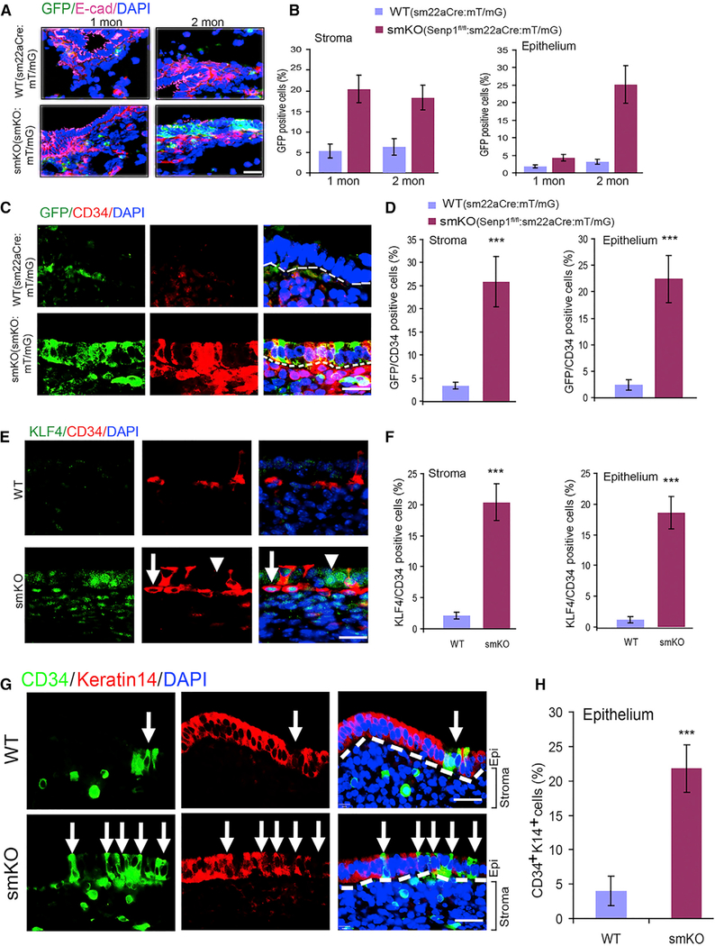
Figure 4. SM22α+CD34+ Stromal Progenitor Cells Directly Contribute to Uterine Hyperplasia.
(A) Immunofluorescent staining with allophycocyanin (APC)-conjugated anti-E-cadherin shown in purple in uterine sections from mT/mG reporter mice (WT) and SENP1smKO:mT/mG mice at the age of 1 and 2 months. DAPI was used for counterstaining of cell nuclei.
(B) GFP+ as indicative SM22α+ cells in the stroma and epithelium layer was quantified.
(C and D) Immunofluorescent staining (C) and quantification (D) of CD34 and co-localization with GFP (SM22α) in uterine sections of 2-month-old WT and SENP1smKO mice.
(E and F) Co-immunofluorescent staining (E) and quantification (F) of KLF4 and CD34 in uterine sections of 2-month-old WT and SENP1smKO mice. Arrow indicates a typical nuclear KLF4-staining CD34+ progenitor cell, while the arrowhead indicates CD34 cells located in the epithelium layer with KLF4-cytoplasmic staining.
(G and H) Co-immunofluorescent staining (G) and quantification (H) of CD43 and epithelial marker keratin 14 in uterine sections of 2-month-old WT and SENP1smKO mice. White dashed lines show the boundaries between the endometrial stroma and epithelium. White arrows show CD34+ keratin 14− cells in the epithelial layer.
All of the data are presented as means ± SEMs; n = 5; **p < 0.01 and ***p < 0.001 (two-sided Student’s t test). Scale bars: 20 μm (A, C, E, and G).

