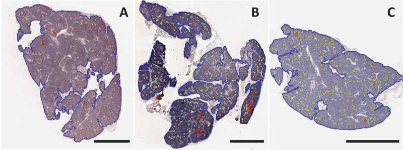
Figure 2.

Image analysis of Congo Red staining for islet amyloid in three patients with type 1 diabetes. Sections were stained and analyzed with Congo Red as described in Methods. Whole slide scans were manually annotated for total pancreas area (blue) and islets (yellow). Amyloid area within islets (red) was identified using the tissue classifier within HALO image analysis software. A pancreas body section from case 6371 (A) and pancreas tail from 6362 (C) showed scattered and infrequent islet amyloidosis. A pancreas tail section from case 6414 (B) showed clustering of islet amyloidosis in two lobules (shown in higher magnification in Figure 3). Scale bars: 2 mm. Whole slide images are available through the nPOD Online Pathology Database (https://www.jdrfnpod.org/for-investigators/online-pathology-information/).
