
FIGURE 10-3.
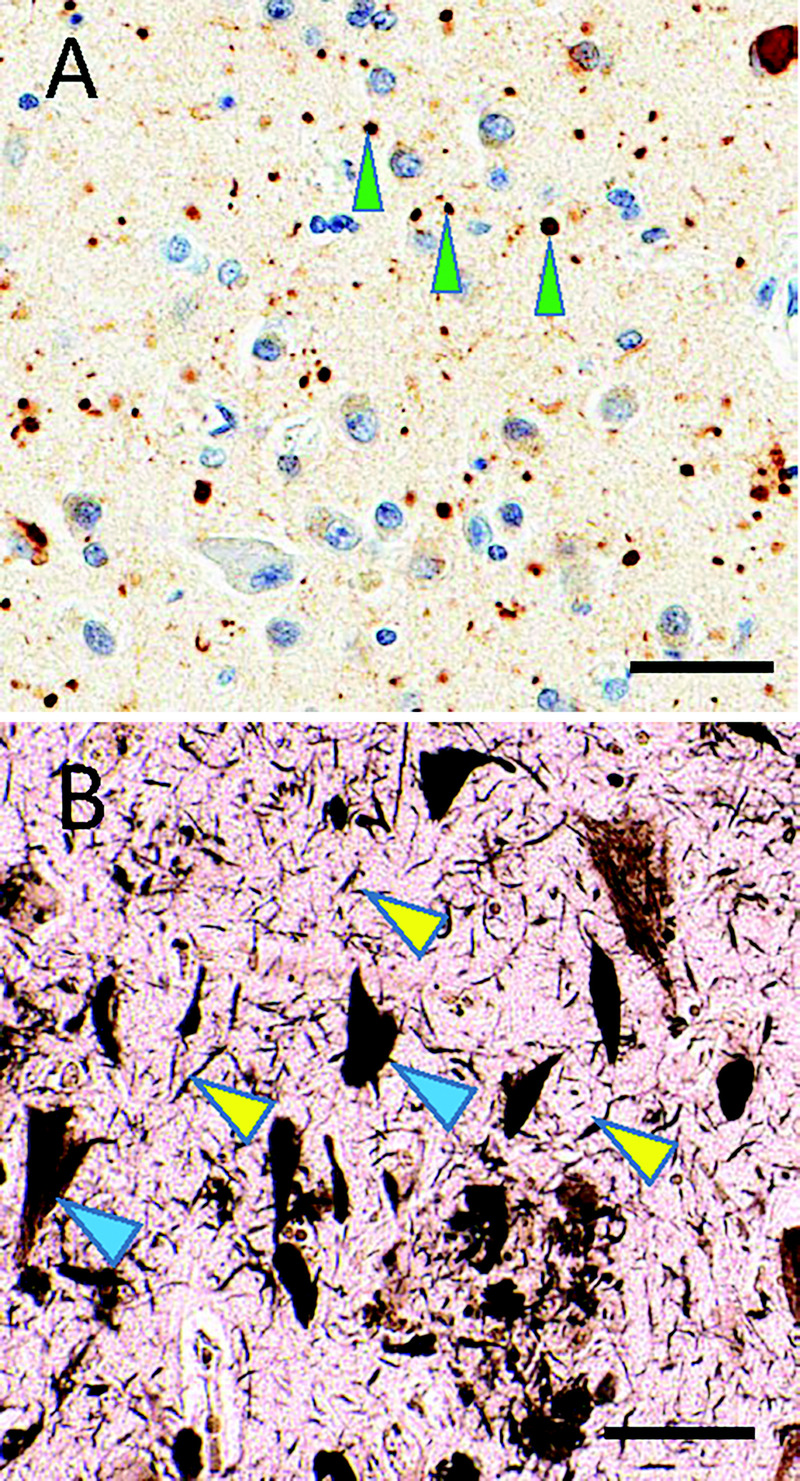
Subtypes of tauopathy. A, Immunostaining shows phosphorylated tau (brown) and counterstaining hematoxylin shows cell nuclei (blue) in argyrophilic grain disease. Note the roundish, approximately 5-micron immunostained structures (green arrowheads), which constitute “grains.” B, In contrast, silver staining highlights Alzheimer disease–type neurofibrillary pathology that includes threadlike structures (yellow arrowheads) and neurofibrillary tangles in neuronal cytoplasm (blue arrowheads). Scale bars: 50 microns in A; 40 microns in B.