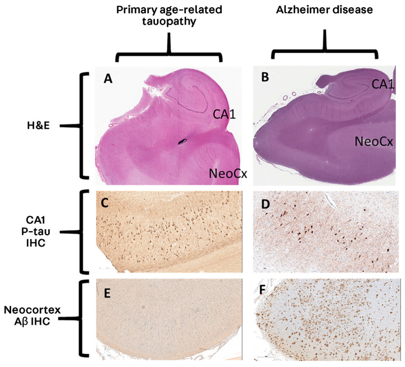
FIGURE 10-4.

Comparison of primary age-related tauopathy and Alzheimer disease. A, B, Analogous sections from hippocampal-level regions stained with hematoxylin and eosin (H&E) at 1× magnification. C, D, Immunohistochemistry for phosphorylated tau (p-tau) at 20× magnification. E, F, Immunohistochemistry for amyloid-β (Aβ) at 10× magnification. Note that the appearances of the stained sections are very similar for p-tau (C, D); by contrast, the neocortical regions lack Aβ in primary age-related tauopathy (E) but show many Aβ plaques in Alzheimer disease (F).
CA1 = cornu ammonis 1; IHC = immunohistochemistry; NeoCx = neocortex.
