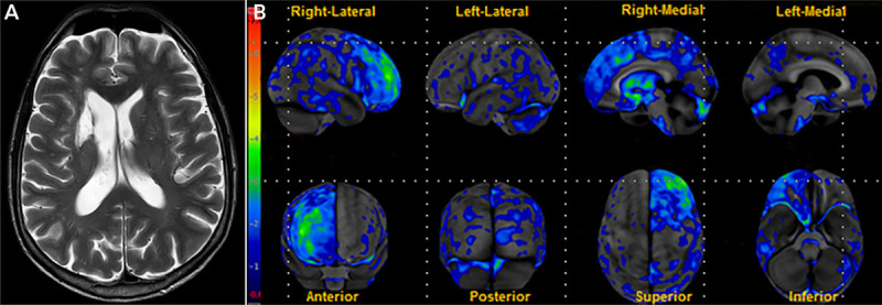
FIGURE 7-5.

Imaging of the patient in case 7-4. A, Axial T2-weighted MRI shows an infarct involving the right caudate. B, Fludeoxyglucose positron emission tomography (FDG-PET) statistical map shows regions of significant hypometabolism relative to age-matched controls. Hypometabolism is present in areas functionally connected to the caudate, including the right medial prefrontal cortex, dorsolateral prefrontal cortex, and right anterior cingulate cortex. Contralateral cerebellar hypometabolism is also seen.
Reprinted with permission from Graff-Radford J, et al, Neurology.50 © 2017 American Academy of Neurology.
