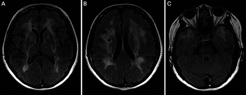
FIGURE 7-7.

Imaging of the patient in case 7-6. Axial fluid-attenuated inversion recovery (FLAIR) MRI shows subcortical ischemic infarcts (A, B) in addition to white-matter T2 hyperintensities with notable involvement of the anterior temporal lobes (C) and external capsule (A) characteristic of cerebral autosomal dominant arteriopathy with subcortical infarcts and leukoencephalopathy (CADASIL).
