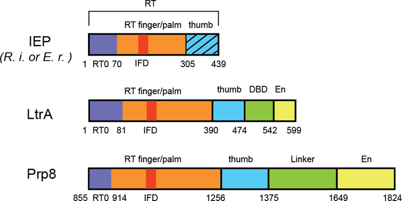
Figure 1.

Comparison of domain architecture of group II IEPs and Prp8. RT, reverse transcriptase domain; DBD, DNA binding domain; EN, endonuclease domain. The hatched pattern in the IEP represents the thumb domain missing in the structures of Zhao and Pyle. Image courtesy of Yaming Shao.
