
Figure 2.
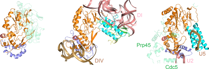
Comparison of RT and RT-like domains from R.i., LtrA and Prp8. Protein domains and motifs are color coded as in Fig. 1. Left: R.i. RT dimer with one protomer colored transparent green. Middle: RNP containing LtrA IEP and group II intron. DI and DIV are colored in salmon and sand, respectively. Right: The RT-like fragment of Prp8 (PDB ID: 3JB9). Interacting proteins are labeled and shown as transparent green; U2, U5 spliceosomal RNA components are shown in salmon and sand, respectively. Image courtesy of Yaming Shao.