
Figure 2.
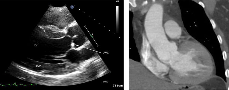
Imaging findings in a 43 year old man who presented with a systolic blood pressure of 290 mmHg and type B aortic dissection. Left panel: Parasternal long axis view on transthoracic echocardiogram of the left ventricle (LV) showing cardiac findings characteristic of chronic hypertension. The septal (S) and posterior wall (PW) thickness is greater than 1.5 cm (referenced to 1 cm-spaced tick marks along the right side of the image), consistent with moderate concentric hypertrophy. There is mild aortic valve calcification (AVC, arrow). Right panel: Coronal view of a chest computed tomography scan from the same patient demonstrating concentric hypertrophy of the left ventricle.