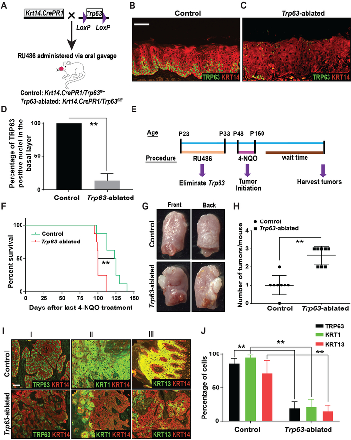
Figure 2. Ablation of Trp63 promotes oral tumorigenesis.
(A) Schematic illustrating the generation of control and Trp63-ablated mice. Immunostaining with antibodies against TRP63 (green) and KRT14 (red) on tongue sections from (B) control and (C) Trp63-ablated mice. TP63 staining studies were performed using the p63α antibody. (D) Quantification of TRP63 positive nuclei from B and C (two-tailed unpaired Student t-test, **: P < 0.01; Mean ± SD). (E) Experimental approach for ablation of Trp63 and generation of oral SCCs by 4-NQO treatment. (F) Graph depicting the survival of control and Trp63-ablated mice (n=8/group) (log-rank test, **: P < 0.01). (G) Representative photographs of SCCs on the front and back of the tongue from control and Trp63-ablated mice. (H) Graph depicting the number of tumors per mouse in control and Trp63-ablated mice (Mann-Whitney U-test, **: P < 0.01) (n=8/group). (I) Immunostaining with antibodies against (I) TRP63 (green) and KRT14 (red), (II) KRT1 (green) and KRT14 (red), and (III) KRT13 (green) and KRT14 (red), on SCCs from control and Trp63-ablated mice. Scale bars = 50 μm. (J) Quantification of TRP63, KRT1 and KRT13 expression from I (two-tailed unpaired Student t-test, **: P < 0.01; Mean ± SD).

