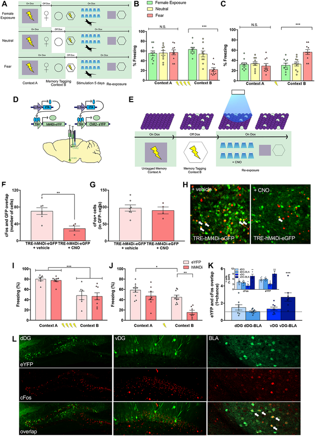
Figure 3. Chronic activation of memories induces long-lasting and bidirectional changes in behavior.
(A) A virus cocktail of AAV9-c-Fos-tTA and AAV9-TRE-ChR2-eYFP was infused into the dorsal and ventral DG. All groups were first fear conditioned in context A, followed by fear conditioning in context B a day later, during which the fear group was off Dox. A separate group was exposed to a neutral context to label active cells with ChR2 and returned to being on Dox. Memories were reactivated twice a day for 5 days in a novel context. (B) Upon re-exposure to Contexts A and B, chronic reactivation of cells processing a four-shock fear memory in the dorsal DG resulted in reduced freezing in the tagged context (e.g. Context B) compared to reactivation of cells processing female and neutral memories (n=10/group. Repeated measures ANOVA, Group × Context, [F(2, 27)=12.67, p<0.01]). Newman-Keules posthoc test revealed significantly lower levels of freezing in Context B compared to Context A only in fear group (p<0.001). (C) When returned to contexts A and B, chronic reactivation of cells processing a single-shock fear memory in the ventral DG resulted in increased freezing in the tagged context (e.g. Context B) compared to reactivation of cells processing female and neutral memories (n=10/group: Repeated measures ANOVA, Group × Context, [F(2, 27)=10.88, p<0.01]). Posthoc revealed significantly higher levels of freezing in Context B compared to Context A only in fear group (p<0.001). (D,E) A virus cocktail of AAV9-c-Fos-tTA and AAV9-TRE-ChR2-eYFP (ChR2) was infused into the dorsal or ventral hippocampus and AAV9-c-Fos-tTA and AAV9-TRE-hM4Di-eYFP (hM4Di) or AAV9-TRE-eYFP (eYFP) was infused into the basolateral amygdala (BLA). Mice were fear conditioned in Context A while on Dox and in Context B while off Dox to label DG and BLA cells associated with Context B fear memory with ChR2 and hM4Di or eYFP, respectively. Following tagging, mice underwent a total of 10 sessions over 5 days of optical stimulation while in a novel environment; all mice received 1 mg/kg CNO (i.p.) 30 minutes prior to each session. Finally, mice were placed back into Context A and Context B and their levels of freezing were assessed (in the absence of either light activation or CNO injection). (F) Treatment with CNO significantly reduced cFos and GFP overlap when compared to treatment with a vehicle control (Student’s t-test, p = 0.006). (G) cFos expression in GFP− cells was comparable between CNO- and vehicle-treated brains following CNO infusion. (H) Representative images of cFos (red) and eGFP (green) overlap in the BLA following CNO treatment. (I) Chronic reactivation of cells processing a strong four-shock fear conditioning memory in the dorsal DG led to a context-specific reduction of freezing behavior, an effect not affected by hM4Di-expression in the BLA. (eYFP: n=6; hM4Di: n=8; Repeated-Measures ANOVA, Main effect of Context [F(1,12)=41.93, p<0.001] without a main effect of Group or Context × Group interaction (ns). (J) Chronic reactivation of cells processing a weak single-shock fear conditioning memory in the ventral DG led to a context-specific increase in freezing behavior. This effect was ablated by chemogenetic inactivation of the BLA (eYFP: n = 10; hM4Di: n = 7; Repeated-Measures ANOVA, Main effect of Context [F(1,15) = 23.92, p < 0.001], Main effect of Group [F(1,15) = 15.47, p < 0.01]). Posthoc revealed significantly lower levels of freezing between the eYFP and hM4Di group in Context A (p < 0.001). (K) Following chronic activation of dDG fear memories, there was no significant change in overlap between eYFP+ and cFos+ cells in either the dDG or BLA (denoted here as dDG-BLA). However, after chronic activation of vDG fear memories, there was a significant amount of overlap between eYFP+ and c-Fos+ cells above chance (dashed line) in the BLA (denoted by vDG-BLA), but not in the vDG. Inset represents % positive cFos+ and eYFP+ cells. cFos+ and eYFP+ cells were significantly greater in the BLA after chronic vDG activation compared to all other groups. (L) Representative images of eYFP (green), c-Fos (red), and overlap in the dDG, vDG, and BLA. White arrows indicate cells expressing eYFP and c-Fos. See also Figures S1 and S2.

