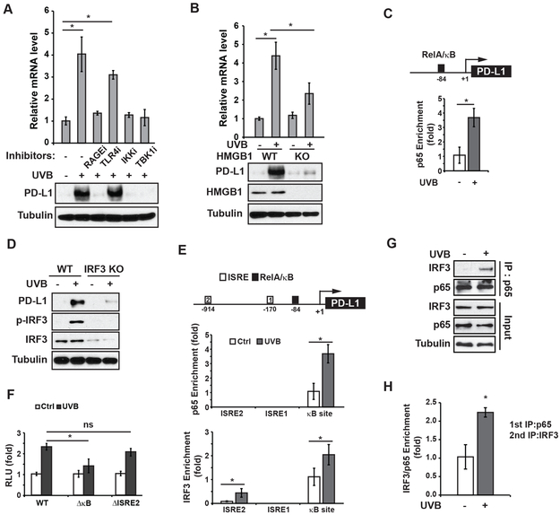
Figure 5. PD-L1 induction by UVB depends on NF-κB and IRF3.
(A) SK-Mel-28 cells were exposed to UVB with or without indicated inhibitors. After 24 h, the expression of PD-L1 was examined by qPCR and western blots. (B) Relative PD-L1 mRNA levels were measured in wild-type (WT) and HMGB1-knockout (KO) SK-Mel-28 cells after UVB exposure. Whole-cell extracts were analyzed by western blots. (C) UVB-induced enrichment of p65 in the PD-L1 promoter region was determined by chromatin immunoprecipitation (ChIP). (D) The PD-L1 expression level was analyzed in WT and IRF3-KO SK-Mel-28 cells after UVB exposure by western blot as in (B). (E) UVB-induced enrichment of p65 and IRF3 on respective ISRE and NF-κB (κB) sites within the PD-L1 promoter region was determined by ChIP. (F) PD-L1 promoter reporters with or without indicated motif deletion were transfected into SK-Mel-28 cells. The UVB-induced reporter activity was measured by luciferase assay. (G) SK-Mel-28 cells were treated with UVB (50 mJ/cm2). Cells were harvested at 4 h after treatment and the interaction of p65 and IRF3 was analyzed with co-IP. (H) ChIP/ReChIP assay was conducted by sequential IP with p65 (1st IP) and IRF3 (2nd IP) antibodies. Enrichment of p65/IRF3 complex on PD-L1 promoter region was determined by qPCR. *: p<0.05, ns: not significant.

