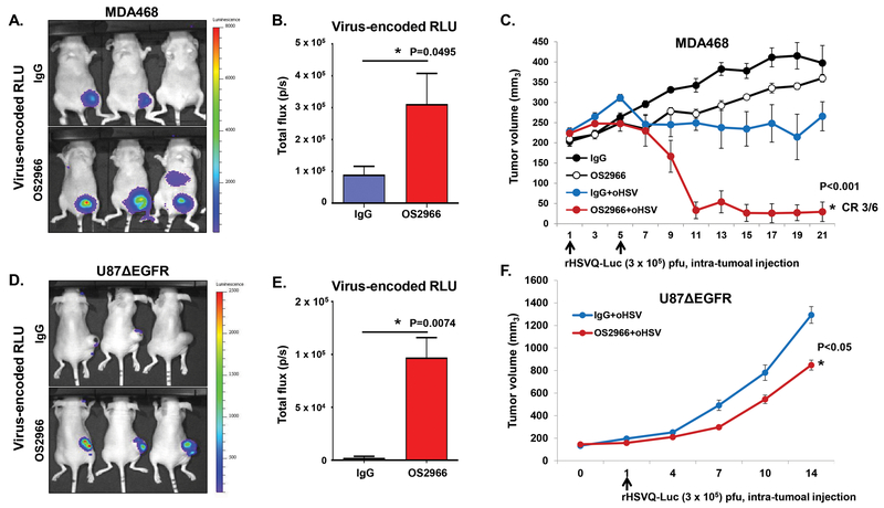
Figure 4. OS2966 treatment increases virus replication and anti-tumor efficacy in vivo.
A-C) Athymic nude mice were implanted with MDA468 by mammary fat pad injection. When tumor size reached around 200 mm3, mice were treated with control IgG or OS2966 (5mg/kg) for 1 day prior to oHSV treatment and twice a week thereafter for the duration of the study, and with 3 × 10⁵ pfu of rHSVQ-Luc on day 1 and 5 (total twice). A) Representative luciferase images of oHSV-treated mice with IgG/OS2966 on day 4. B) Data shown are total flux in each mouse, indicating quantification of virally expressed luciferase gene activity in MDA468 mammary fat pad injected mice with IgG or OS2966 on day 4. C) Tumor volume was measured regularly after treatment and data shown are the mean tumor volumes ± SEM, at the indicated time points (n = 6/group). * indicates p < 0.05; rHSVQ-Luc+OS2999 versus rHSVQ-Luc + IgG. D-F) Athymic nude mice were subcutaneously implanted with U87ΔEGFR. When tumor size reached around 150 mm3, 1 × 10⁵ pfu of an oHSV expressing luciferase (rHSVQ-Luc) were injected intratumorally. Control human IgG or OS2966 (5 mg/kg) were administered intraperitoneally on days −1, 1, 3, and 5 after oHSV therapy. D) Representative luciferase images of oHSV-treated mice with IgG/OS2966 on day 3. E) Data shown are total flux in each mouse treated with IgG or OS2966 on day 3. F) Tumor volume was measured regularly after treatment and data points represent the mean of the tumor volumes ± SEM, at the indicated time points (n = 6/group). * indicates p < 0.05; rHSVQ-Luc+OS2999 versus rHSVQ-Luc + IgG.

