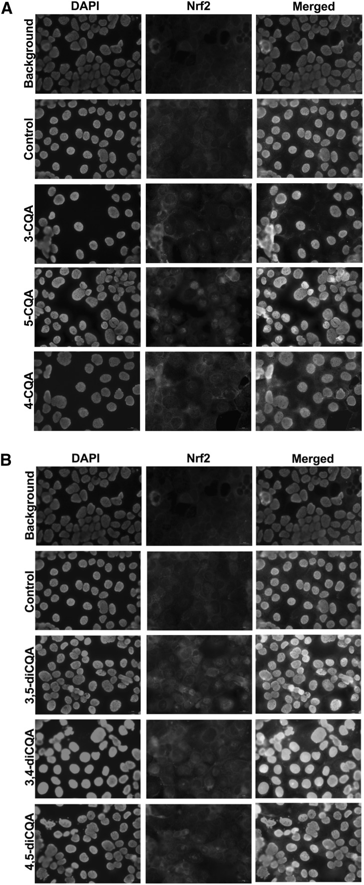
Fig. 3.
Immunocytochemistry of cells showing different individual CGA isomers at 0.2 mM enhanced Nrf2 localization into nucleus in Caco-2 cells. a 3-CQA, 5-CQA, and 4-CQA. b 3,5-diCQA, 3,4-diCQA, and 4,5-diCQA Localization of Nrf2 was performed by double immunofluorescence staining in cells with only PMA + IFNγ treatment, cells with individual CGA isomer treatment before PMA + IFNγ challenge. Background represents cells treated with PMA + IFNγ but without a primary antibody during the immunofluorescence staining. The control represents cells treated with PMA + IFNγ, and with both primary and secondary antibodies during the immunofluorescence staining. Nrf2 protein were stained in green; nuclei were stained with DAPI (blue). The merged image showed the nuclear location of Nrf2 in nuclei. (Color figure online)

