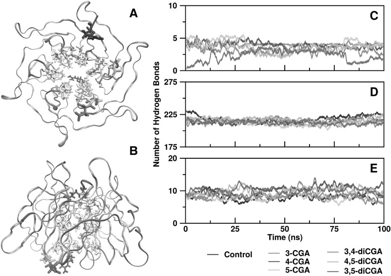
Fig. 5.
Analysis of hydrogen bonds in CGA isomers that interacted with Keap1/Nrf2 complex. Bottom (a) and side (b) views of Keap1 β-propeller according to residue types. Blue, arginine; red, aspartic acid; green, glycine, serine, or threonine; white, valine, alanine, isoleucine or leucine. Hydrogen bonds between CGA isomer and Keap1 β-propeller (c) and intra-Keap1 β-propeller (d), and between Keap1 β-propeller and Nrf2 fragment (e). (Color figure online)

